Featured Software, Algorithms, and Code


Interactive Calculator
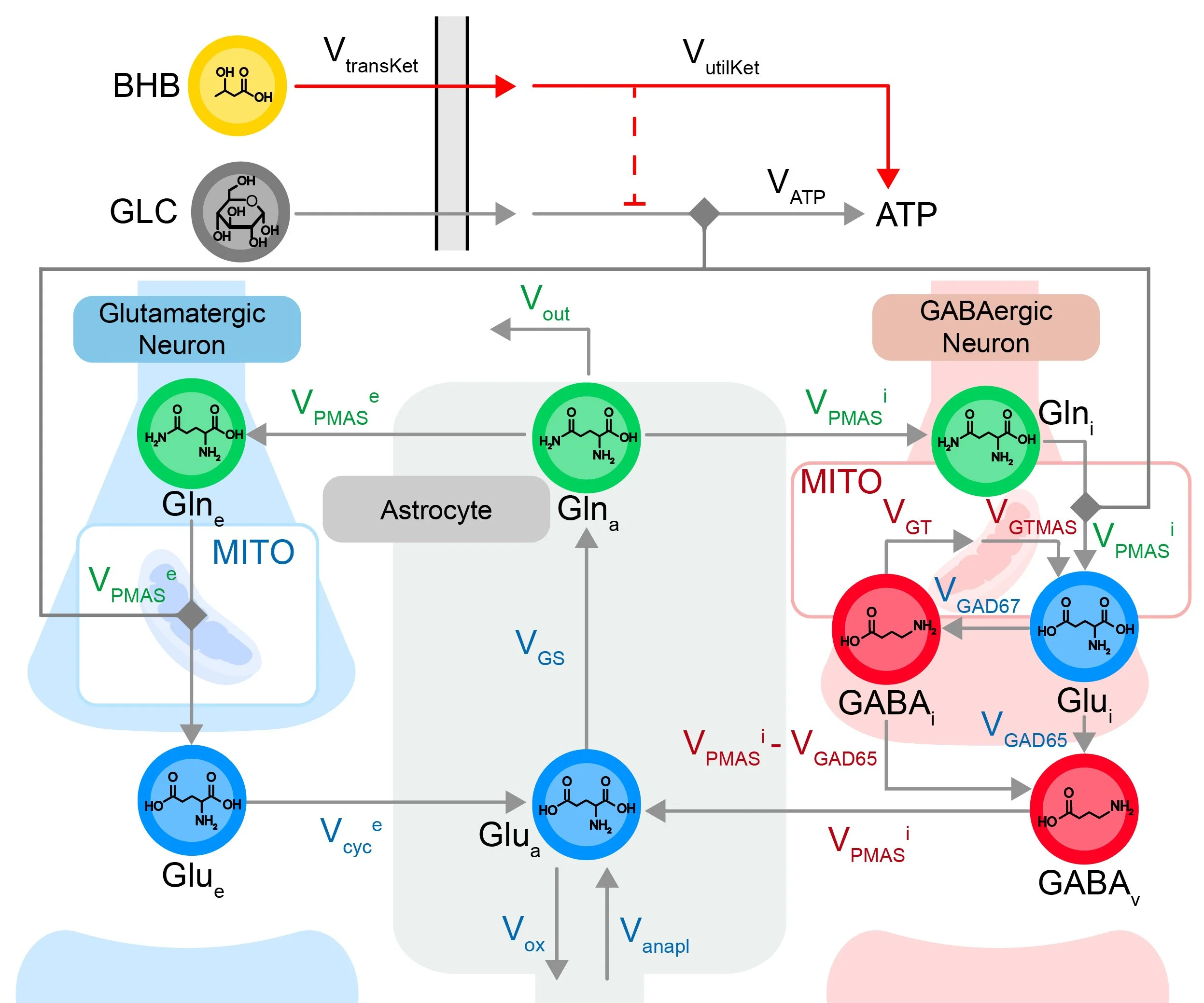
Ketones vs. Glucose Neurotransmitter Cycling

-
Although computational models have deepened our understanding of neuroscience, it is still highly challenging to link actual low-level physiological activity (spiking, field potentials) and biochemistry (transmitters and receptors) directly with high-level cognitive abilities (decision-making, working memory) and associated disorders. Here, we introduce a mechanistically accurate multi-scale model directly generating simulated physiology from which extended neural and cognitive phenomena emerge. The model produces spiking, fields, phase synchronies, and synaptic change, directly generating working memory, decisions, and categorization. These were then validated on extensive experimental macaque data from which the model received no prior training of any kind. Moreover, the simulation uncovered a previously unknown neural code (“incongruent neurons”) that specifically predicts upcoming erroneous behaviors, also subsequently confirmed in empirical data. The biomimetic model thus directly and predictively links decision and reinforcement signals, of computational interest, with spiking and field codes, of neurobiological importance.
Read article in Nature Communications.
-
All fields of science depend on mathematical models. Occam's razor refers to the principle that good models should exclude parameters beyond those minimally required to describe the systems they represent. This is because redundancy can lead to incorrect estimates of model parameters from data, and thus inaccurate or ambiguous conclusions. Here, we show how deep learning can be powerfully leveraged to address Occam's razor. FixFit, our new method, uses a feedforward deep neural network with a bottleneck layer to characterize and predict the behavior of a given model from its input parameters. FixFit has three major benefits. First, it provides a metric to quantify the original model's degree of complexity. Second, it allows for the unique fitting of data. Third, it provides an unbiased way to discriminate between experimental hypotheses that add value versus those that do not. In two use cases, we demonstrate the broad applicability of this method across scientific domains. To validate the method using a known system, we apply FixFit to recover known composite parameters for the Kepler orbit model. To illustrate how the method can be applied to less well-established fields, we use it to identify parameters for a multi-scale brain model and reduce the search space for viable candidate mechanisms.
Read article in Computational Biology.
-
Using neuroimaging and electrophysiological data to infer neural parameter estimations from theoretical circuits requires solving the inverse problem. Here, we provide a new Julia language package designed to i) compose complex dynamical models in a simple and modular way with ModelingToolkit.jl, ii) implement parameter fitting based on spectral dynamic causal modeling (sDCM) using the Laplace approximation, analogous to MATLAB implementation in SPM12, and iii) leverage Julia’s unique strengths to increase accuracy and speed by employing Automatic Differentiation during the fitting procedure. To illustrate the utility of our flexible modular approach, we provide a method to improve correction for fMRI scanner field strengths (1.5T, 3T, 7T) when fitting models to real data.
Read article in Imaging Neuroscience.
-
As a field, control systems engineering has developed quantitative methods to characterize the regulation of systems or processes, whose functioning is ubiquitous within synthetic systems. In this context, a control circuit is objectively “well regulated” when discrepancy between desired and achieved output trajectories is minimized and “robust” to the degree that it can regulate well in response to a wide range of stimuli. Most psychiatric disorders are assumed to reflect dysregulation of brain circuits. Yet, probing circuit regulation requires fundamentally different analytic strategies than the correlations relied upon for analyses of connectivity and their resultant networks. Here, we demonstrate how well-established methods for system identification in control systems engineering may be applied to functional magnetic resonance imaging (fMRI) data to extract generative computational models of human brain circuits. As required for clinical neurodiagnostics, we show these models to be extractable even at the level of the single subject. Control parameters provide two quantitative measures of direct relevance for psychiatric disorders: a circuit’s sensitivity to external perturbation and its dysregulation.
Read Article in Computational Brain & Behavior
Neuroblox beta version and tutorials now available through MIT OpenCourseWare.
Neuroblox Metabolic Module

Neuroblox is a new computational neuroscience platform optimized for probing the behavior of neural control circuits and their regulation. Neuroblox is explicitly data-driven, multi-scale (spiking-neuron, LFP, M/EEG, fMRI) and coded for high-performance computing in Julia with a user-friendly graphical user interface. Neuroblox is designed to allow neuroscientists to identify, simulate, and estimate quantitative parameters for these control circuits in response to different stimuli, disorders, and therapeutics.
Featured Instrumentation Development
BrainDancer is the first commercially available dynamic phantom for fMRI, designed to markedly increase signal/noise and dynamic fidelity of fMRI time series. Applications include data-driven optimization of acquisition parameters, superior artifact removal, and normalization across sessions, scanners, and sites for longitudinal and multi-site studies.
Introduction to BrainDancer
Setting Up BrainDancer
Analyzing Your Data with BrainDancer
-
The fMRI community has made great strides in decoupling neuronal activity from other physiologically induced T2* changes, using sensors that provide a ground-truth with respect to cardiac, respiratory, and head movement dynamics. However, blood oxygenation level-dependent (BOLD) time-series dynamics are also confounded by scanner artifacts, in complex ways that can vary not only between scanners but even, for the same scanner, between sessions. Unfortunately, the lack of an equivalent ground truth for BOLD time-series has thus far stymied the development of reliable methods for identification and removal of scanner-induced noise, a problem that we have previously shown to severely impact detection sensitivity of resting-state brain networks. To address this problem, we first designed and built a phantom capable of providing dynamic signals equivalent to that of the resting-state brain. Using the dynamic phantom, we then compared the ground-truth time-series with its measured fMRI data. Using these, we introduce data-quality metrics: Standardized Signal-to-Noise Ratio (ST-SNR) and Dynamic Fidelity that, unlike currently used measures such as temporal SNR (tSNR), can be directly compared across scanners. Dynamic phantom data acquired from four “best-case” scenarios: high-performance scanners with MR-physicist-optimized acquisition protocols, still showed scanner instability/multiplicative noise contributions of about 6–18% of the total noise. We further measured strong non-linearity in the fMRI response for all scanners, ranging between 8–19% of total voxels. To correct scanner distortion of fMRI time-series dynamics at a single-subject level, we trained a convolutional neural network (CNN) on paired sets of measured vs. ground-truth data. The CNN learned the unique features of each session's noise, providing a customized temporal filter. Tests on dynamic phantom time-series showed a 4- to 7-fold increase in ST-SNR and about 40–70% increase in Dynamic Fidelity after denoising, with CNN denoising outperforming both the temporal bandpass filtering and denoising using Marchenko-Pastur principal component analysis. Critically, we observed that the CNN temporal denoising pushes ST-SNR to a regime where signal power is higher than that of noise (ST-SNR > 1). Denoising human-data with ground-truth-trained CNN, in turn, showed markedly increased detection sensitivity of resting-state networks. These were visible even at the level of the single-subject, as required for clinical applications of fMRI.
-
As the only FDA-approved near-infrared fluorophore, indocyanine green (ICG) is commonly used to image vasculature in vivo. ICG degrades rapidly in solution, which limits its usefulness in certain applications, including time-sensitive surgical procedures. We propose formulations that address this shortcoming via complexation with β-cyclodextrin derivatives (β-CyD), which are known to create stabilizing inclusion complexes with hydrophobic molecules. Here, we complexed ICG with highly soluble methyl β-CyD and FDA-approved sulfobutyl ether β-CyD (Captisol®) in aqueous solution. We measured the fluorescence of the complexes over 24 h. We found that both CyD+ICG complexes exhibit sustained fluorescence increases of >2.0× versus ICG in water and >20.0× in PBS. Using transmission electron microscopy, we found evidence of reduced aggregation in complexes versus ICG alone. We thus conclude that this reduction in aggregation helps mitigate fluorescence autoquenching of CyD+ICG complexes compared in ICG alone. We also found that while ICG complexed with methyl β-CyD severely reduced the viability of MRC-5 fibroblasts, ICG complexed with sulfobutyl ether β-CyD had no effect on viability. These results represent an important first step toward enhancing the utility of aqueous ICG by reducing aggregation-dependent fluorescence degradation. © 2015
Read Article in J Biomed Mater Res Part B: Appl Biomater
-
Substance abuse is a fundamentally dynamic disease, characterized by repeated oscillation between craving, drug self-administration, reward, and satiety. To model nicotine addiction as a control system, a magnetic resonance imaging (MRI)-compatible nicotine delivery system is needed to elicit cyclical cravings. Using a concentric nebulizer, inserted into one nostril, we delivered each dose equivalent to a single cigarette puff by a syringe pump. A control mechanism permits dual modes: one delivers puffs on a fixed interval programmed by researchers; with the other, subjects press a button to self-administer each nicotine dose. We tested the viability of this delivery method for studying the brain's response to nicotine addiction in three steps. First, we established the pharmacokinetics of nicotine delivery, using a dosing scheme designed to gradually achieve saturation. Second, we lengthened the time between microdoses to elicit craving cycles, using both fixed-interval and subject-driven behavior. Finally, we demonstrate a potential application of our device by showing that a fixed-interval protocol can reliably identify neuromodulatory targets for pharmacotherapy or brain stimulation. Our MRI-compatible nasal delivery method enables the measurement of neural circuit responses to drug doses on a single-subject level, allowing the development of data-driven predictive models to quantify individual dysregulations of the reward control circuit causing addiction.
Read Article in Pharmaceutics